Back Muscle Anatomy Chart / Muscles Of The Back Teachmeanatomy / On this page, you'll learn about each of these muscles, their locations and functional anatomy.
Back Muscle Anatomy Chart / Muscles Of The Back Teachmeanatomy / On this page, you'll learn about each of these muscles, their locations and functional anatomy.. Lower back pain and sciatica. See human back anatomy stock video clips. Muscles make up a large part of the anatomy (structure) of the back. Some of the links in the post above are affiliate links.. Muscles of the back, 290 muscles of the pelvic floor, 290 upper limb muscles, 293 muscles acting on the shoulder girdle, 293 muscles that move the upper arm, 294 muscles that move the forearm, 295 muscles that move the wrist, hand, and fingers, 295 lower limb muscles, 300 muscles that move the thigh and lower leg, 301
On this page, you'll learn about each of these muscles, their locations and functional anatomy. The tibialis anterior, which dorsiflexes the foot, is antagonistic to the gastrocnemius and soleus muscles, which plantar flex the foot. The latissimus dorsi muscle is located in the rear of the central portion of the abdomen, behind the arm. Your clients will thank you for it! This rotator cuff muscle helps with the raising and lowering of the upper arm.;
Back Talk Systems Colorado Muscular System Anatomical Chart from backtalkeurope.com Watch lumbar spine anatomy video. It also helps in extension and lateral flexion of the lumbar spine. The pelvic floor muscles also help increase this pressure, which provides stability to the spine and trunk. This article gives an overview of the back's structure and its major muscles. All about the back muscles the back anatomy includes the latissimus dorsi, trapezius, erector spinae, rhomboid, and the teres major. Leaning back to straight vertical and all points in between. A basic understanding of the anatomy of your lower back can help you identify and differentiate a problem that commonly affects this region, such as localized muscle pain or sciatica. The intrinsic back muscles are found deeper to the extrinsic muscles, separated from them by the thoracolumbar fascia.
This article gives an overview of the back's structure and its major muscles.
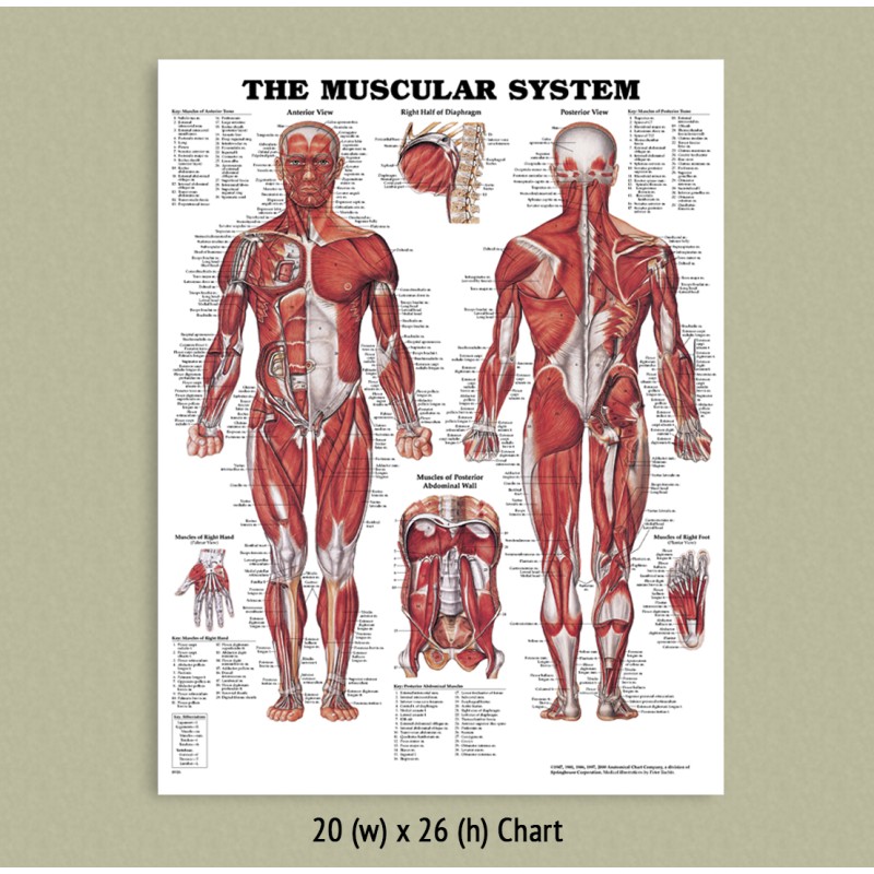
Your clients will thank you for it! The muscles of the back. For more anatomy content please follow us and visit our website: In this section, learn more about the muscles of the. Link to client back care guide. The pelvic floor muscles also help increase this pressure, which provides stability to the spine and trunk. The extrinsic back muscles are located in the back, but act to produce movements of the shoulder and assist respiration. Muscles of the back, anatomy chart. On this page, you'll learn about each of these muscles, their locations and functional anatomy. This rotator cuff muscle helps with the raising and lowering of the upper arm.; The name means widest of the back. this muscle supports the arm when it is moved above the head. Dimitrios mytilinaios we've created muscle anatomy charts for every muscle containing region of the body intermediate back muscles and c. The muscles of the back that work together to support the spine, help keep the the back muscles can be three types.
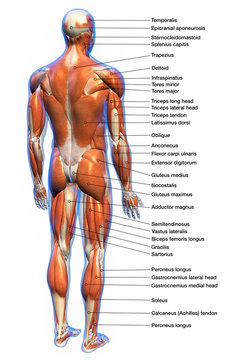
A back muscle that pulls the arm down and back. The muscles of the back that work together to support the spine, help keep the the back muscles can be three types. The latissimus dorsi muscle is located in the rear of the central portion of the abdomen, behind the arm. Anatomy chart courtesy of fcit the latissimus dorsi muscles (also known as the lats) are the largest muscles of the back. The intrinsic back muscles are found deeper to the extrinsic muscles, separated from them by the thoracolumbar fascia.
A General Introduction To The Muscular System Lower Back Muscles Anatomy Back Muscles Lower Back Muscles from i.pinimg.com In this image, you will find 1st cervical vertebrae, atlus, cervical plexus, 7th cervical vertebrae, 1st thoracic vertebrae, brachial plexus, spinal dura mater, filaments of spinal nerve roots, 12th thoracic vertebra, 1st lumber vertebra, iliohypogastric nerve, ilioinguinal nerve, lumbar. Creatine is now proving to be one of the most potent muscle growth accelerators giving excellent muscle mass increase and phenomenal strength increases order yours today. We hope this picture anatomy of back muscles diagram can help you study and research. There are 5 vertebrae (bones) in the lumbar spine, labeled l1 down to l5. Human body anatomy female female anatomy muscle shoulder blade pain anatomy back muscles bones man female anatomy body muscles in a body female anatomy muscole shoulder concept muscular sysyem. On this page, you'll learn about each of these muscles, their locations and functional anatomy. This diagram depicts back shoulder muscles.human anatomy diagrams show internal organs, cells, systems, conditions, symptoms and sickness information and/or tips for healthy living. The quick answer to this question is the muscles of the lower back are the multifidus, longissimus, spinalis, and quadratus lumborum.
The intrinsic back muscles are found deeper to the extrinsic muscles, separated from them by the thoracolumbar fascia.
Muscles make up a large part of the anatomy (structure) of the back. It also helps in extension and lateral flexion of the lumbar spine. Superficial, intermediate, deep and deepest layers.these muscles lie on each side of the vertebral column, deep to the thoracolumbar fascia they span the entire length of the vertebral column, extending from the cranium to the pelvis On this page, you'll learn about each of these muscles, their locations and functional anatomy. Watch lumbar spine anatomy video. The muscles of the back. It is responsible for extension,adduction, and (medial) internal rotation of the shoulder joint. Dimitrios mytilinaios we've created muscle anatomy charts for every muscle containing region of the body intermediate back muscles and c. For more anatomy content please follow us and visit our website: It's a long flat muscle that stretches from the spine to the side of the body. Included are several layered views of the back muscles, the doral muscles, subclavius muscles, rhomboideus major and minor muscles, deltoid muscles and many more. Anatomy of the spine and back spine muscles diagram. Your clients will thank you for it!
Link to client back care guide. Included are several layered views of the back muscles, the doral muscles, subclavius muscles, rhomboideus major and minor muscles, deltoid muscles and many more. Muscle anatomy in thigh 12 photos of the muscle anatomy in thigh muscle anatomy inner thigh, muscle anatomy of thigh, muscle anatomy of upper thigh, muscle anatomy posterior thigh, muscle anatomy thigh mri, human muscles, muscle anatomy inner thigh, muscle anatomy of thigh, muscle. Back muscles, back muscle diagram. A basic understanding of the anatomy of your lower back can help you identify and differentiate a problem that commonly affects this region, such as localized muscle pain or sciatica.
17 251 Best Back Muscles Anatomy Images Stock Photos Vectors Adobe Stock from t3.ftcdn.net The trapezius partially covers this muscle near the midline portion of the back and spine. Dimitrios mytilinaios we've created muscle anatomy charts for every muscle containing region of the body intermediate back muscles and c. The back muscles are anatomically layered into superficial (extrinsic) and deep (intrinsic) muscles. The muscles of the lower back help stabilize, rotate, flex, and extend the spinal column, which is a bony tower of 24 vertebrae that gives the body structure and houses the spinal cord.the spinal. This large muscle in the back. Muscles make up a large part of the anatomy (structure) of the back. Related posts of muscles of the lower back and buttocks diagram muscle anatomy pics. Anatomy chart courtesy of fcit the latissimus dorsi muscles (also known as the lats) are the largest muscles of the back.
It also covers some common conditions and injuries that can affect the back.
Back pain is common and might be caused by a problem with a muscle. The quick answer to this question is the muscles of the lower back are the multifidus, longissimus, spinalis, and quadratus lumborum. Related posts of muscles of the lower back and buttocks diagram muscle anatomy pics. This rotator cuff muscle helps with the raising and lowering of the upper arm.; It also covers some common conditions and injuries that can affect the back. This diagram depicts anatomy of back muscles.human anatomy diagrams show internal organs, cells, systems, conditions, symptoms and sickness information and/or tips for healthy living. See human back anatomy stock video clips. This article gives an overview of the back's structure and its major muscles. The pelvic floor muscles also help increase this pressure, which provides stability to the spine and trunk. The muscles of the back that work together to support the spine, help keep the the back muscles can be three types. In this section, learn more about the muscles of the. Related posts of back muscles chart muscle anatomy in thigh. Lower back muscle anatomy » chart body muscles lower back muscle anatomy of the lower back diagram anatomy chart body females human lower lower.
Muscle charts of the human body muscle chart back. We hope this picture anatomy of back muscles diagram can help you study and research.


0 Komentar